
Menopause May Be the First Alzheimer’s Warning
Women face higher Alzheimer’s risk, and menopause may be the turning point. Discover what research says about symptoms, hormones, and prevention.

Women have been told for decades the same story about Alzheimer’s: it’s a disease of old age, and women are affected more often simply because they live longer.
That explanation no longer holds.
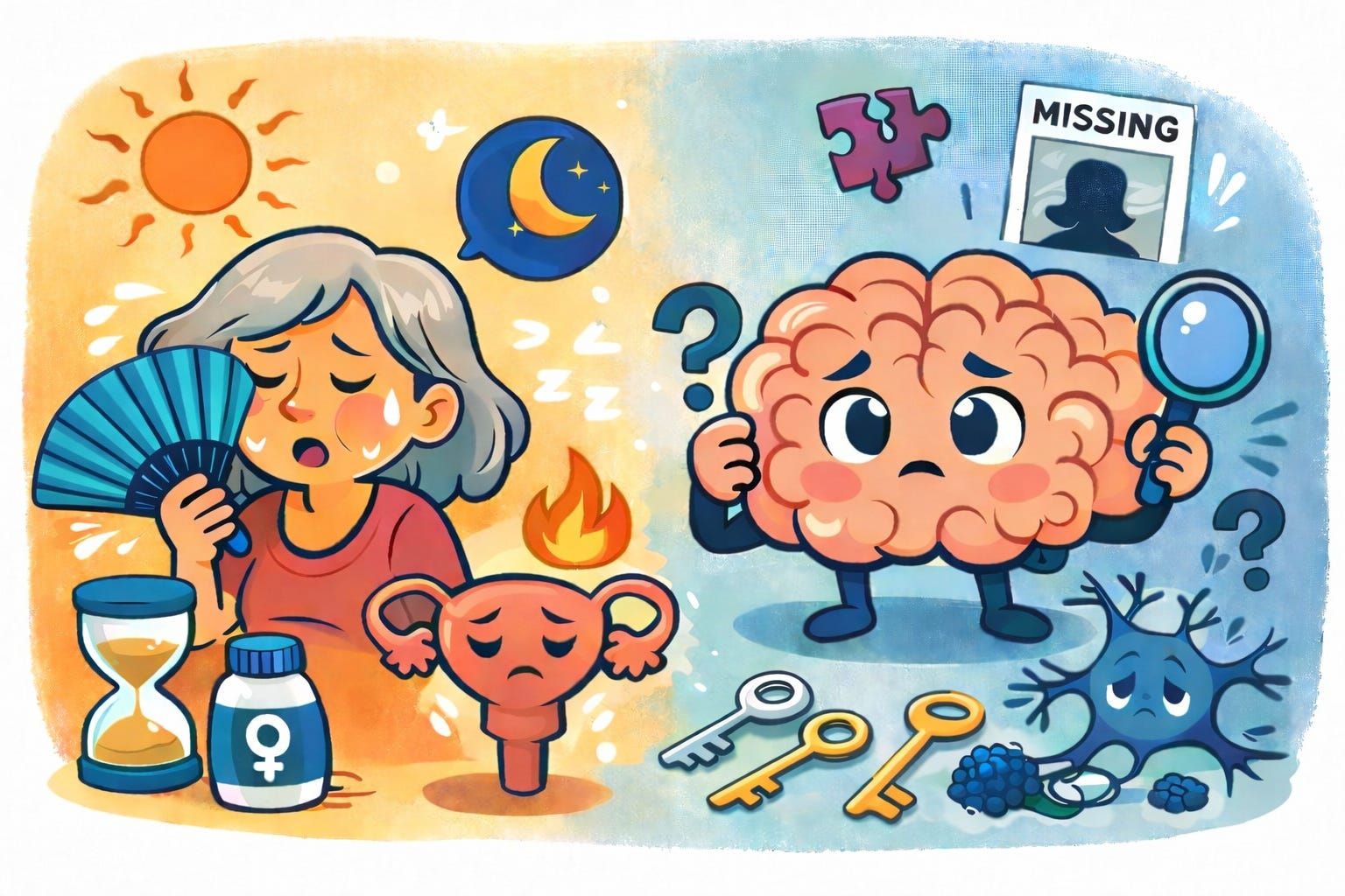
The data now point to a different story: women are not just outliving men into Alzheimer’s, they are more biologically vulnerable to it. And the earliest changes may begin not in old age, but in midlife, during the menopausal transition, when symptoms like brain fog, insomnia, anxiety, and memory lapses are still being dismissed as stress or hormones.
By the time a woman is told to worry about Alzheimer’s, her brain may have been asking for attention for years.
Two-Thirds of Alzheimer’s Patients Are Women
7.2 million Americans are living with Alzheimer’s disease.
Nearly 4.8 million of them are women.
The explanation medicine has leaned, that women simply live longer and Alzheimer’s is a disease of longevity, has been systematically dismantled by neuroimaging data collected over the past decade.
Longevity accounts for some of the disparity, but it does not account for most of it.
A landmark study in Neurology examined lifetime risk trajectories in 5,000 participants and found that at age 45, a woman’s lifetime risk of developing Alzheimer’s is 1 in 5. A man’s is 1 in 10.1 When researchers controlled for survival, the disparity did not disappear. Women face a fundamentally higher biological vulnerability, independent of how long they live.
Alzheimer’s Begins in the Menopausal Transition
The conventional model of Alzheimer’s placed the disease firmly in late life: a problem of the 70s and 80s, triggered by aging. Brain imaging has forced a revision of that model.
Dr. Lisa Mosconi’s research group at Weill Cornell Medicine has spent over a decade scanning the brains of women across the menopausal transition. Their findings are consistent and striking: the structural and metabolic changes associated with Alzheimer’s risk begin during perimenopause, not post-menopause, and not in old age.2
The 2021 multi-modal neuroimaging study, which examined PET, MRI, and functional connectivity data in 161 women, found that the transition from pre- to post-menopause was associated with measurable decreases in gray matter volume in memory-critical regions, reductions in mitochondrial ATP production, and increased amyloid-β deposition.3 These changes tracked with menopausal endocrine status, not chronological age.
“Hot flashes, insomnia, brain fog, depression, anxiety: those symptoms don’t start in the ovaries. They start in the brain. Those are neurological symptoms.” — Dr. Lisa Mosconi
Brain fog in a 44-year-old woman is often dismissed as stress, sleep deprivation, or perimenopause. It may be all three simultaneously. It may also be the earliest detectable signal of a neurological process that, left unaddressed, will not plateau.
When Your Brain Loses Its Architect
Estrogen is not a reproductive hormone that occasionally visits the brain. Estrogen receptors are expressed throughout the hippocampus, prefrontal cortex, and limbic system, the same regions responsible for memory, executive function, and emotional regulation.
Estrogen supports multiple neuroprotective functions: it modulates inflammation, supports mitochondrial efficiency, maintains the cholinergic neurons that produce acetylcholine (a neurotransmitter essential for memory consolidation), and regulates the microglial cells that serve as the brain’s immune system.
When estrogen begins its erratic perimenopausal decline, several of these systems shift simultaneously. Microglia can move toward a pro-inflammatory activated profile. Cholinergic neurons, deprived of estrogenic support, may reduce acetylcholine output. These are not theoretical mechanisms extrapolated from rodent studies. They are measurable changes on PET and MRI imaging in living human women.3
Symptoms that begin during perimenopause, including word-finding difficulty, mood instability, disrupted sleep, and memory lapses, are not peripheral complaints to be managed with reassurance.
The Hormone No One Is Measuring
There is a second biological story unfolding in parallel with the estrogen narrative, and it receives far less attention: cortisol.
A 2018 study in Neurology, using data from the Framingham Heart Study (2,231 dementia-free participants, mean age 48.5), found that individuals in their 40s with the highest morning cortisol levels performed significantly worse on measures of memory, visual perception, and executive function.4 The sex-specific finding was striking: the association between elevated cortisol and reduced total brain volume was significant only in women, not in men.
A subsequent study extended these findings, showing that in women, elevated cortisol was associated with higher beta-amyloid accumulation in Alzheimer’s-vulnerable brain regions.5
The women most at risk are often the ones medicine considers its most capable patients: high-functioning, high-responsibility, managing everything. The chronic cortisol load of that life is neurotoxic, and it’s not being assessed.
We Forgot to Worry About This
For decades, FSH, follicle-stimulating hormone, was understood as a messenger: a signal the pituitary sends to the ovaries when it wants them to produce more estrogen. Clinicians used it as a diagnostic marker. They did not think of it as a risk factor.
A 2022 study in Nature demonstrated in preclinical models that FSH binds directly to hippocampal and cortical neurons to accelerate both amyloid-β and tau deposition.6 A 2023 study from Mosconi’s group showed that in living women, higher FSH levels were independently associated with greater amyloid-β load in the frontal cortex.7
A 2025 Mendelian randomization study found a significant causal association between genetically elevated FSH levels and Alzheimer’s disease risk.8 A woman whose FSH is rising, even while she is still cycling, may already be on a neurological trajectory that no one is tracking.
The Appointment That Doesn’t Happen
The standard model, screening for cognitive decline in the 70s and 80s, is built around a disease that, in women, may have been progressing for two decades by the time any screen catches it.
A baseline cognitive assessment in a woman’s mid-40s, combined with FSH levels, morning cortisol, APOE genotyping, and metabolic markers, does not diagnose dementia. It establishes a reference point. It creates the ability to detect drift. It moves the clinical conversation from reactive to preventive.
Three questions worth asking at your next appointment:
What is my FSH level, and is it being tracked longitudinally?
Can we establish a baseline cognitive assessment this year?
Has anyone measured my morning cortisol or APOE genotype?
The paid post includes the lab panel to request at midlife, FSH thresholds, HRT evidence, the supplement stack, and the 7 precision interventions with the strongest mechanistic evidence.
Subscribe to download The Alzheimer Prevention Protocol.